Medical Thermography

Breast
Thermography

Specific Area
Thermography

Breast Thermography + Ultrasound

Full Body
Thermography

Half Body
Thermography

Full Body Thermography + Ultrasound
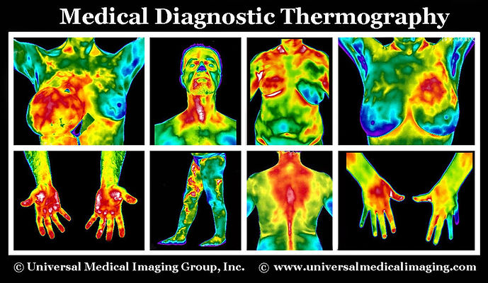
Thermography is an adjunctive state-of-the art Medical Diagnostic Infrared Thermal Imaging which can detect physiological abnormalities by measuring temperature changes. An infrared scanning device is used to convert infrared signal emitted from the skin surface into electrical impulses that are seen in color on a monitor. This visual image graphically maps the body temperature and is referred to as a thermogram.
Thermography’s major clinical value is in its high sensitivity to pathology in the vascular, muscular, neural and skeletal systems and as such can contribute to the pathogenesis and diagnosis made by the clinician. It is a life saving procedure that can alert you and your doctor to changes in your body that may indicate early stage breast disease or other disease processes that may be developing in the body. Thermography offers the opportunity of earlier detection of breast abnormalities than has been possible through breast self examination, doctor examination, or other anatomical tests alone and without any pain, radiation, or compression.
By performing thermography years before conventional mammography, a selected patient population at risk can be monitored more carefully, and then by accurately utilize ultrasound as soon as is possible to detect the actual lesion - (once it has grown large enough and dense enough to be seen), can increase the patients treatment options and ultimately improve the outcome. It is in this role that thermography provides it’s most practical benefit to the general public and to the medical profession. It is certainly an adjunct to the appropriate usage of other diagnostic medical tests and not a competitor.
Thermography is a completely non-invasive, without radiation, painless clinical imaging procedure for detecting and monitoring a number of diseases and physical injuries by showing thermal abnormalities present in the body. It is used as an aid for diagnosis and prognosis, as well as monitoring therapy progress, for conditions and injuries, including:
Back Injuries
Arthritis
Headache
Nerve Damage
Unexplained Pain
Fibromyalgia
RSD (CRPS)
Dental and TMJ
Artery Inflammation
Vascular Disease
Breast Disease
Breast Cancer
Carpal Tunnel Syndrome
Disc Disease
Inflammatory Pain
Skin Cancer
Referred Pain Syndrome
Sprain / Strain
Stroke Screening
Digestive Disorders
X-Ray, C.T., Ultrasound and M.R.I are all tests of 'anatomy' that measure the structures of your body whereas thermography is unique in its capability to show the physiological change and metabolic processes occurring in the body. Although many people associate thermography as a breast screening, images can be taken of the entire body or just regions of interest. The digitized images are stored on a computer and are sent electronically for interpretations, analysis and report. Your report is sent directly to you and to any healthcare professionals requested.
Thermography for Breast Cancer Prevention
Early Detection Saves Lives!
Current research has determined that the key to breast cancer survival rests upon its earliest possible detection. If it’s discovered in its earliest stages, 95% cure rates are possible.
Breast self-examination involves checking the breasts to help detect breast problems or changes. Many breast problems are first discovered by women themselves, often by accident. Breast self-examination involves checking the breasts for lumps or changes while standing and lying in different positions and while looking at the breasts in a mirror to note any changes in their appearance. Once a woman knows what her breasts normally look and feel like, any new lump or change in appearance should be evaluated by a doctor. Breast lumps can be noncancerous (benign) or cancerous (malignant).
In its early stages, breast cancer usually has no symptoms. As a tumor develops, you may note the following signs:
-
A lump in the breast or underarm that persists after the menstrual cycle. This is often the first apparent symptom of breast cancer. Lumps associated with breast cancer are usually painless, although some may cause a noticeable sensation. Lumps are usually visible on a diagnostic medical ultrasound long before they can be visually seen or felt.
-
Swelling in the armpit.
-
Redness, pain or tenderness in the breast. Although lumps are usually painless, pain or tenderness can be a sign of breast cancer.
-
A noticeable flattening or indentation on the breast, which may indicate a tumor that cannot be seen or felt.
-
Any change in the size, contour, texture, or temperature of the breast. A reddish, pitted surface like the skin of an orange could be a sign of advanced breast cancer.
-
A change in the nipple, such as a nipple retraction, dimpling, itching, a burning sensation, or ulceration.
-
Unusual discharge from the nipple that may be clear, bloody or another color. It's usually caused by benign conditions but could be due to cancer in some cases.
-
A marble-like area under the skin.
-
An area that is distinctly different from any other area on either breast.
If breast symptoms and/or the results of your physical exam suggest breast cancer might be present, more tests will probably be ordered by your doctor. These might include different imaging tests. The safest, painless, non-invasive, affordable breast screening tests are a combination of an Ultrasound and Thermography, which may give us about 95% accuracy in detecting breast cancer.
Studies have shown that by the time a tumor has grown to sufficient size to be detectable by physical examination or mammography, it has in fact been growing for about seven years achieving more than 25 doublings of the malignant cell colony. At 90 days there are two cells, at one year there are 16 cells, and at five years there are 1,048,576 cells--an amount that is still undetectable by a mammogram. Thermography has the ability to provide women with future risk assessment. If discovered, certain thermographic risk markers can warn a woman that she needs to work closely with her doctor with regular checkups to monitor her breast health.
If Breast Thermography (test of physiology) combined with Breast Ultrasound (test of anatomy) can help to discover abnormality and also breast cancer in its earliest stage. These safe diagnostic tests can be done on early bases for a regular check up, or more often if the problem was detected, to monitor a treatment progress.
Please remember -- early detection, which includes self examination and safe, painless, non-invasive Ultrasound and Thermography screenings with NO radiation Saves Lives!
Breast Ductal Cell Carcinoma Case Study
______________________________________________________________________________
For more information or to make an appointment for a Comprehensive Full Body Screening please contact
Universal Medical Imaging Group at 818-508-8895 or universalmedicalimaging@yahoo.com